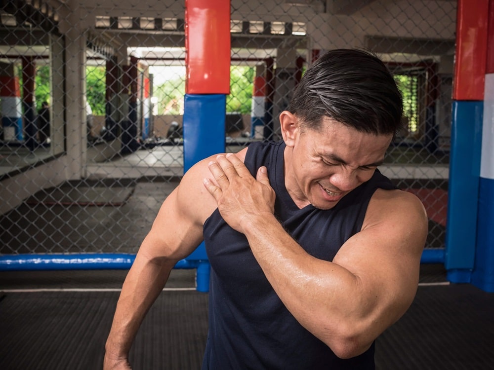
Shoulder arthroscopy surgery is a minimally invasive surgical procedure used to diagnose and treat various shoulder conditions, including shoulder instability, rotator cuff tears, labral tears, impingement syndrome, and shoulder arthritis.
It involves using a small camera called an arthroscope and specialized instruments inserted through tiny incisions in the shoulder to visualize and access the joint’s internal structures.
Here’s an overview of the shoulder arthroscopy procedure:
Preparation: Before surgery, the patient undergoes a preoperative evaluation, including physical examination, imaging studies (such as X-rays, MRI, or CT scans), and a medical history review. The patient may be instructed to refrain from eating or drinking for a certain period before the procedure.
Anesthesia: Shoulder arthroscopy can be performed under general anesthesia, regional anesthesia (such as a nerve block), or local anesthesia with sedation, depending on the patient’s preference and the surgeon’s recommendation.
Incisions: Small incisions, typically less than half an inch in length, are made around the shoulder joint to insert the arthroscope and surgical instruments. These incisions are strategically placed to minimize damage to surrounding tissues and optimize joint visualization.
Arthroscopic Examination: The arthroscope, a thin, flexible tube with a camera and light source at the tip, is inserted through one of the incisions and guided into the shoulder joint. The camera allows the surgeon to visualize the structures inside the joint, including the cartilage, ligaments, tendons, and bony structures.
Surgical Treatment: Once the problem areas in the shoulder joint are identified, Dr. Pournaras will use specialized arthroscopic instruments to perform the necessary surgical procedures. This may include repairing torn ligaments or tendons, removing damaged tissue or bone spurs, debriding (cleaning up) frayed or damaged tissue, or performing other corrective procedures.
Closure: After the surgical procedures are completed, the arthroscope and instruments are removed from the shoulder joint, and the incisions are closed with sutures or surgical tape. Sterile dressings may be applied to the incisions.

Recovery: Following shoulder arthroscopy, the patient is monitored in the recovery area before being discharged home or to a hospital room.
Pain management medications are provided as needed, and instructions are given for wound care, activity restrictions, and rehabilitation exercises.
Rehabilitation: Physical therapy and rehabilitation are typically initiated shortly after surgery to help restore strength, range of motion, and function to the shoulder joint. The specific rehabilitation program may vary depending on the type of surgery performed and the individual patient’s needs.